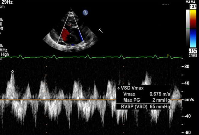
Pulsed-wave tissue Doppler at the tricuspid level of the lateral
By A Mystery Man Writer
Last updated 09 Jul 2024


A Test in Context: E/A and E/e′ to Assess Diastolic Dysfunction

A) An oesophagogastroduodenoscopy 6 weeks postoperative showing

A) An oesophagogastroduodenoscopy 6 weeks postoperative showing

JaypeeDigital

Tuberculoid giant-cellular lesion with giant atypical cells: small

Serdar BOZYEL, Assistant Professor

Pulsed-wave tissue Doppler at the tricuspid level of the lateral

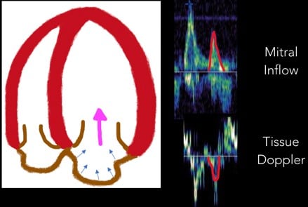
Assessment of diastolic function by echocardiography
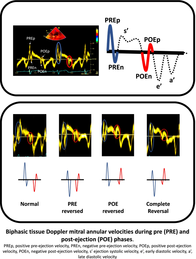
Tissue Doppler derived biphasic velocities during the pre and post
Fellows' Section: Diastology at the Bedside

Tracing derived from pulsed wave tissue Doppler with the sample

A,B: Axial sections, portal phase of abdominal computed tomography
Echocardiography: an overview - Part III

A) Microscopic histology showing focal submucosal invasion, with
Recommended for you
 5mm (3/16 inch) Large Black Pupil 3D Holographic Fishing Lure Eyes, Fl – Crawdads Fishing Tackle14 Jul 2023
 How to make holographic 3d eyes for fishing lures. Free eyes14 Jul 2023
 24 TEARDROP RED SILVER 5mm or 3/16 3D Soft Molded Adhesive Lure14 Jul 2023
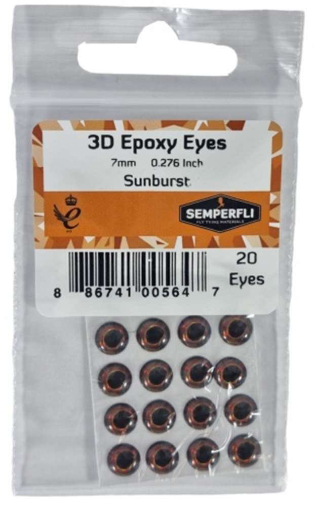 7mm 3D Epoxy Eyes Sunburst14 Jul 2023
 Fishing Lure Eyes,500pcs/pack 5mm 3D Fishing Lure Eye Great Accessory for Make Fishing Bait(red,5mm)14 Jul 2023
 Fishing Lure Eye Orange 3D Soft Eye Fishing Eyes Lure14 Jul 2023
 032 Pop Jr14 Jul 2023
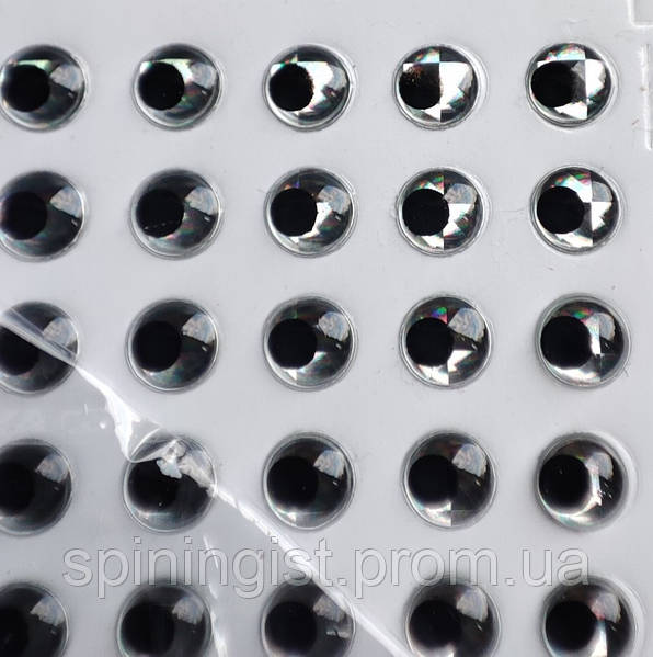 Глазки 3-Д 6 мм (серебренные): продажа, цена в Луцке. Воблеры от14 Jul 2023
 5mm 3D Iridescent #1 / 500 Soft Molded 3D Holographic Fish Eyes, Fly Tying, Jig, Lure Making14 Jul 2023
 Tiyuyo 100pcs Fish Eyes 3D Holographic Lure Eyes Fly Tying Jigs14 Jul 2023
You may also like
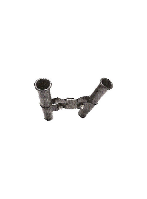 Cannon Dual Front Mount Rod Holders - Marine General14 Jul 2023
- Summer Chic Boutique14 Jul 2023
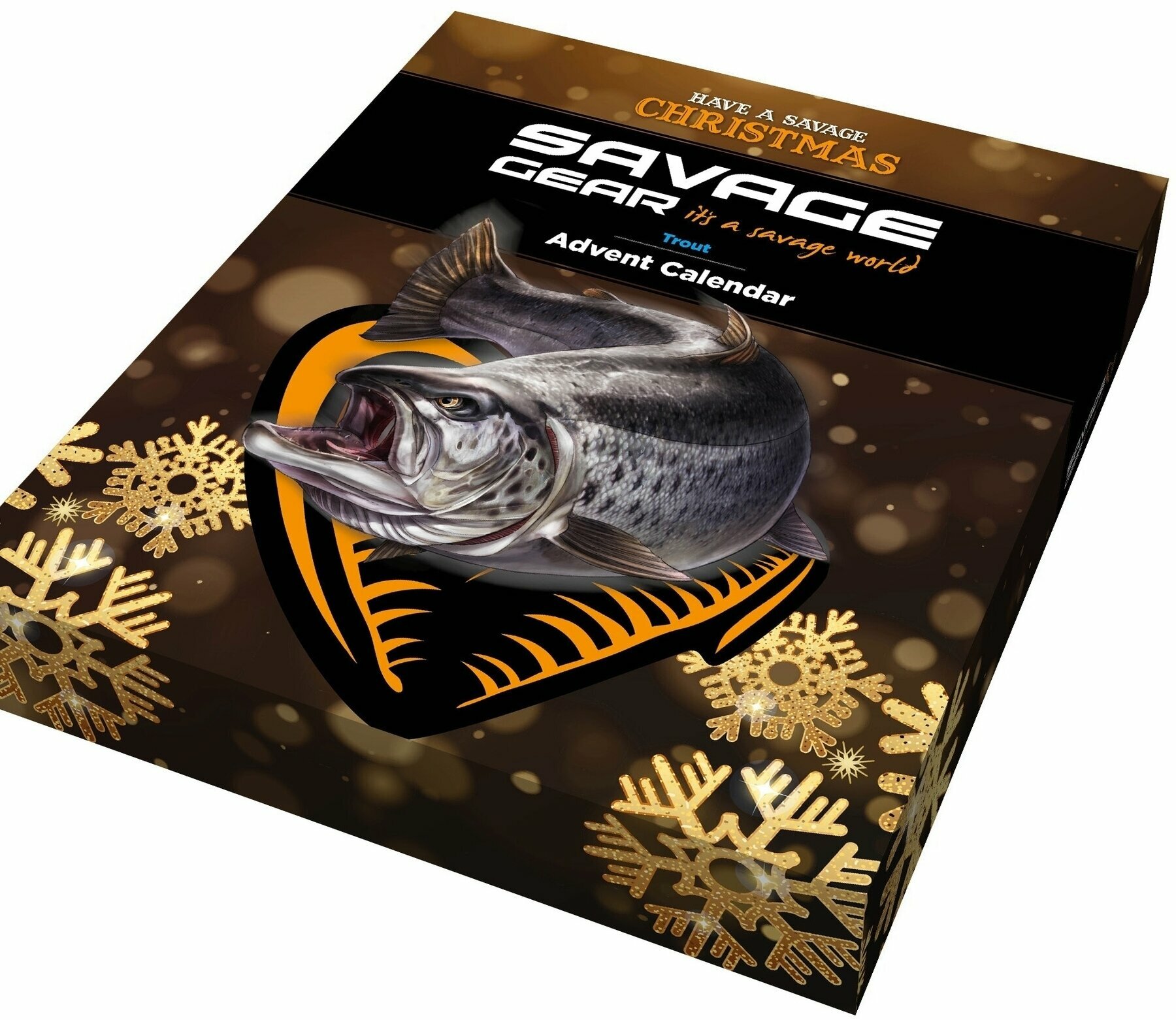 Savage Gear Advent Calendar Seatrout - Muziker14 Jul 2023
 The inside of galvanized water pipe : r/Plumbing14 Jul 2023
 Dive deep into the Bay of Fundy without leaving home14 Jul 2023
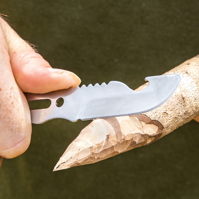 Trailblazer Multi-Function Survival Utensil14 Jul 2023
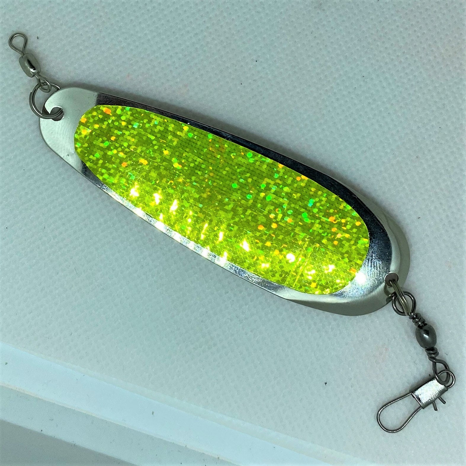 Creekside Mini Wobbler Double Chartreuse Flash14 Jul 2023
 American Girl Doll Size Fidget Spinner - DIY Mini Fidget Spinner without14 Jul 2023
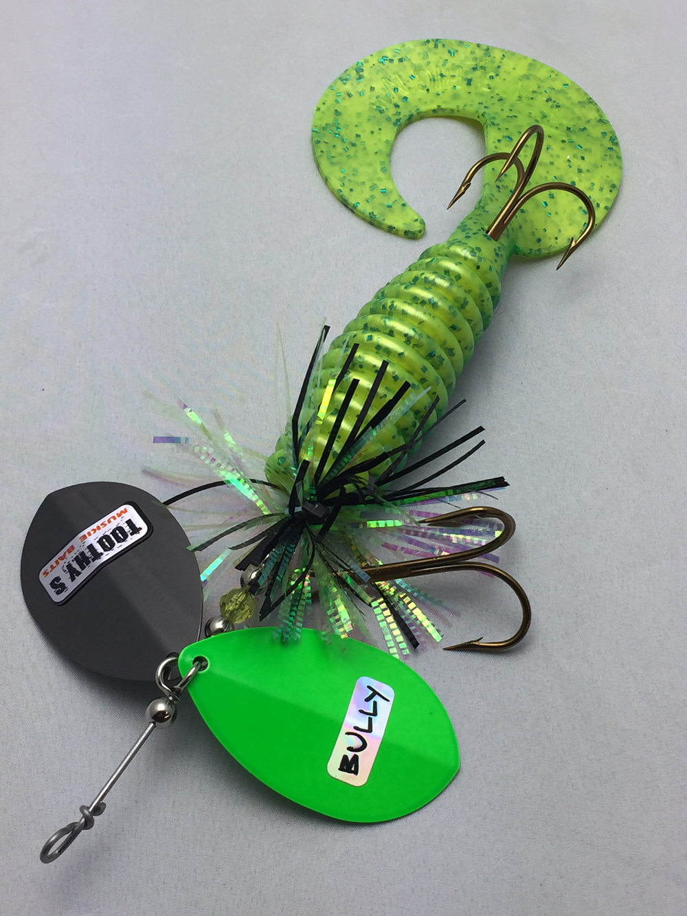 Toothy's Tackle, Musky Lures14 Jul 2023
 Wall reel hozelock auto reel 40m hose koppl - wall hose h14 Jul 2023
